
- #FEATURES OSIRIX MD SOFTWARE#
- #FEATURES OSIRIX MD CODE#
- #FEATURES OSIRIX MD SERIES#
- #FEATURES OSIRIX MD FREE#

#FEATURES OSIRIX MD FREE#
Education downloads - OsiriX MD by Pixmeo and many more programs are available for instant and free download.
#FEATURES OSIRIX MD SERIES#
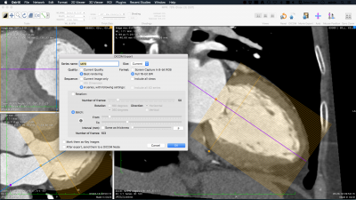
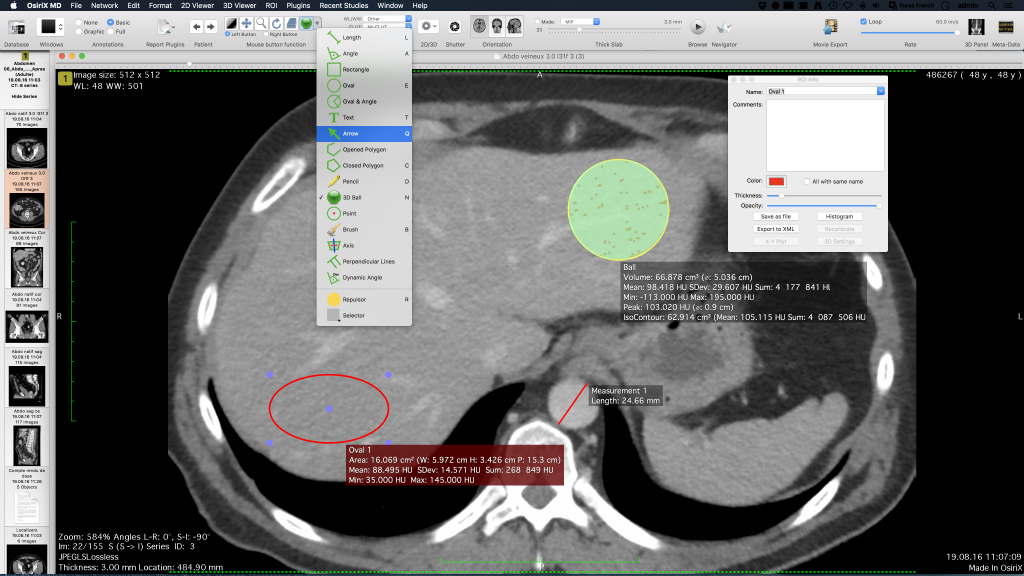
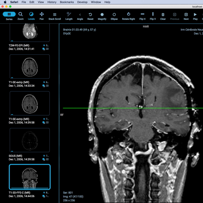
OsiriX has been specifically designed for navigation and visualization of multimodality and multidimensional images: 2D Viewer, 3D Viewer, 4D Viewer (3D series with temporal dimension, for example: Cardiac-CT) and 5D Viewer (3D series with temporal and functional dimensions, for example: Cardiac-PET-CT). OsiriX 11.0 is fully optimized for macOS Catalina 10.15. Initial investigations into the diagnostic capabilities of OsiriX mobile on the iPhone and iPod platforms 6, 7 support this hypothesis. 102(3):127–131, 2001.Ĭopen, W.A., Schaefer, P.W., and Wu, O., MR perfusion imaging in acute ischemic stroke. Laghi, A., Catalano, C., Iannaccone, R., Paolantonio, P., Panebianco, V., Sansoni, I., Trenna, S., and Passariello, R., Multislice spiral CT angiography in the evaluation of the anatomy of splanchnic vessels: Preliminary experience. Jellison, B.J., Field, A.F., Medow, J., Lazar, M., Salamat, M.S., and Alexander, A.L., Diffusion tensor imaging of cerebral white matter: A pictorial review of phisics, fiber tract anatomy, and tumor imaging patterns. Golby, A.J., Kindlmann, G., Norton, I., Yarmarkovich, A., Pieper, S., and Kikinis, R., Interactive diffusion tensor tractography visualization for neurosurgical planning. Kikinis, R., and Pieper, S., 3D slicer as a tool interactive brain tumor segmentation.

17(6):756–759, 2010.įedorov, A., Beichel, R., Kalpathy-Cramer, J., Finet, J., Fillion-Robin, J.C., Pujol, S., Bauer, C., Jennings, D., Fennessy, F., Sonka, M., Buatti, J., Aylward, S., Miller, J.V., Pieper, S., and Kikinis, R., 3D slicer as an image computing platform for the quantitative imaging network. Neuroradiology Report J of Clinical Neurocience.
#FEATURES OSIRIX MD SOFTWARE#
Yamauchi, T., Yamazi, M., Okawa, A., Furuya, T., Hayashi, K., Sakuma, T., Takahashi, H., Yanagawa, N., and Koda, M., Efficacy and reliability of highly functional open source DICOM software (OsiriX) in spine surgery. Sierra, M.E., Cienfuegos, M.R., and Fernández, S.G., OsiriX, a useful tool for processing tomographic images in patients with facial fracture. Lo Presti, G., Carbone, M., Ciriaci, D., Aramiri, D., Ferrari, M., and Ferrari, V., Assesment of DICOM viewers capable of loading patient-specific 3D models obtaines by different segmentation platforms in the operating room. Valeri, G., Mazza, F.A., Maggi, S., Aramini, D., La Riccia, L., Mazzoni, G., and Giovagnoni, A., Open source software in a practical approach for post processing of radiologic images. Karopka, T., Schmuhl, H., and Demski, H., Free/libre open source software in health care: A review. Juanes, J.A., Ruisoto, P., Prats, A., and Framiñán, A., Open source applications for image visualization and processing in neuroimaging training. We will compare the features of the VITREA2® and AW VolumeShare 5® radiology workstation with free open source software applications like OsiriX® and 3D Slicer®, with examples from specific studies. With this study, we aim to present the advantages and disadvantages of these radiological image visualization systems in the advanced management of radiological studies. Nevertheless, the programs included in these workstations have a high cost which always depends on the software provider and is always subject to its norms and requirements. These radiological devices are basically CT (Computerised Tomography), MRI (Magnetic Resonance Imaging), PET (Positron Emission Tomography), etc. In radiology, graphic workstations allow their users to process, review, analyse, communicate and exchange multidimensional digital images acquired with different image-capturing radiological devices. Consequently, sophisticated computing tools that combine software and hardware to process medical images are needed. For the radiological field, manipulating and post-processing images is increasingly important. However, this last group of free applications have limitations in its use. Two examples of free open source software are Osirix Lite® and 3D Slicer®.

#FEATURES OSIRIX MD CODE#
In addition, some of these applications are known as Free Open Source Software because they are free and the source code is freely available, and therefore it can be easily obtained even on personal computers. These software applications are of great interest, both from a teaching and a radiological perspective. Currently, there are sophisticated applications that make it possible to visualize medical images and even to manipulate them.


 0 kommentar(er)
